6. Muscles of the Pectoral Girdle and Upper Limbs
Muscles of the shoulder and upper limb can be divided into four groups: muscles that stabilize and position the pectoral girdle, muscles that move the arm, muscles that move the forearm, and muscles that move the wrists, hands, and fingers.
Muscles That Position the Pectoral Girdle
The pectoral girdle, or shoulder girdle, consists of the lateral ends of the clavicle and scapula, along with the proximal end of the humerus, and the muscles covering these three bones to stabilize the shoulder joint. The girdle creates a base from which the head of the humerus, in its ball-and-socket joint with the glenoid fossa of the scapula, can move the arm in multiple directions.Muscles that position the pectoral girdle are located either on the anterior thorax or on the posterior thorax (Figure 6.1 and Table 11.8). The anterior muscles include the subclavius, pectoralis minor, and serratus anterior. The posterior muscles include the trapezius, rhomboid major, and rhomboid minor. When the rhomboids are contracted, your scapula moves medially, which can pull the shoulder and upper limb posteriorly.
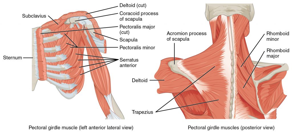
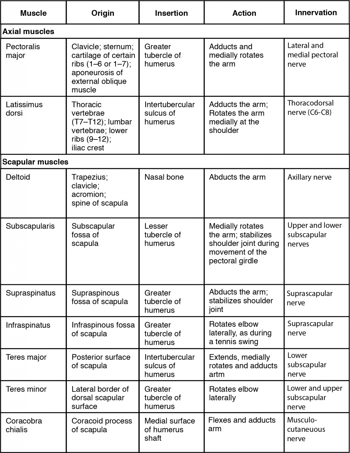
| Muscles that Position the Pectoral Girdle (Table 11.8) | ||||||
|---|---|---|---|---|---|---|
| Position in the thorax | Movement | Target | Target motion direction | Prime mover | Origin | Insertion |
| Anterior thorax | Stabilizes clavicle during movement by depressing it | Clavicle | Depression | Subclavius | First rib | Inferior surface of clavicle |
| Anterior thorax | Rotates shoulder anteriorly (throwing motion); assists with inhalation | Scapula; ribs | Scapula: depresses; ribs: elevates | Pectoralis minor | Anterior surfaces of certain ribs (2–4 or 3–5) | Coracoid process of scapula |
| Anterior thorax | Moves arm from side of body to front of body; assists with inhalation | Scapula; ribs | Scapula: protracts; ribs: elevates | Serratus anterior | Muscle slips from certain ribs (1–8 or 1–9) | Anterior surface of vertebral border of scapula |
| Posterior thorax | Elevates shoulders (shrugging); pulls shoulder blades together; tilts head backwards | Scapula; cervical spine | Scapula: rotests inferiorly, retracts, elevates, and depresses; spine: extends | Trapezius | Skull; vertebral column | Acromion and spine of scapula; clavicle |
| Posterior thorax | Stabilizes scapula during pectoral girdle movement | Scapula | Retracts; rotates inferiorly | Rhomboid major | Thoracic vertebrae (T2–T5) | Medial border of scapula |
| Posterior thorax | Stabilizes scapula during pectoral girdle movement | Scapula | Retracts; rotates inferiorly | Rhomboid minor | Cervical and thoracic vertebrae (C7 and T1) | Medial border of scapula |
Muscles That Move the Humerus
Similar to the muscles that position the pectoral girdle, muscles that cross the shoulder joint and move the humerus bone of the arm include both axial and scapular muscles (Figure 6.2 and Figure 6.3). The two axial muscles are the pectoralis major and the latissimus dorsi. The pectoralis major is thick and fan-shaped, covering much of the superior portion of the anterior thorax. The broad, triangular latissimus dorsi is located on the inferior part of the back and has multiple points of origin including the lumbosacral fascia attached to the inferior 6 thoracic vertebrae, the inferior 3 ribs, the iliac crest and inferior angle of the scapula.
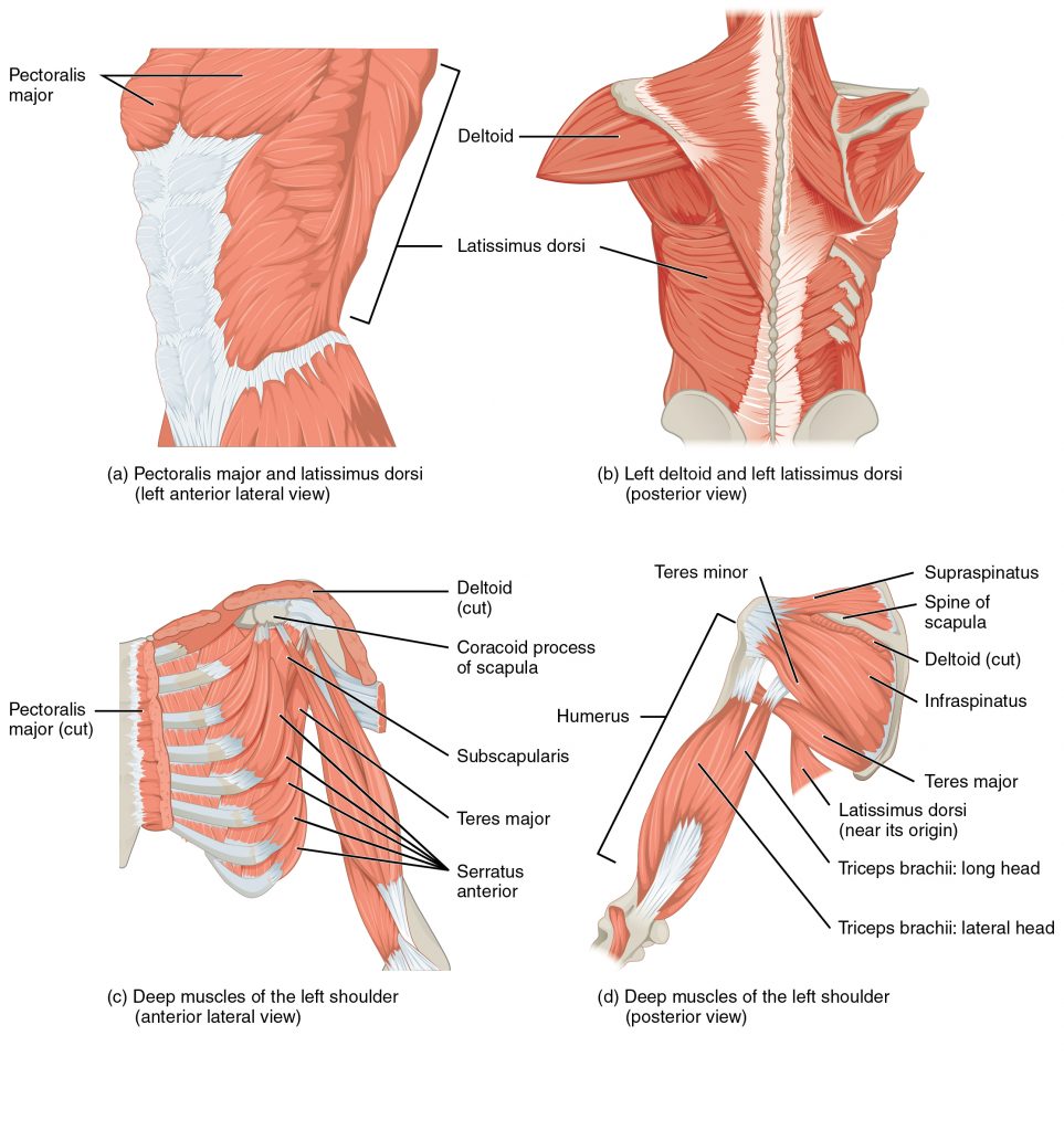
The rest of the shoulder muscles originate on the scapula and help to move the arm. The deltoid is the major abductor of the arm but also facilitates flexing and medial rotation, as well as extension and lateral rotation. The subscapularis originates on subscapular fossa and medially rotates the arm. Named for their locations, the supraspinatus (originating from the supraspinous fossa) and the infraspinatus (originating from the infraspinous fossa) abduct the arm, and laterally rotate the arm, respectively. The thick and flat teres major is inferior to the teres minor and extends the arm, and assists in its adduction and medial rotation. The long teres minor laterally rotates the arm. Finally, the coracobrachialis flexes and adducts the arm.
The tendons of the subscapularis, supraspinatus, infraspinatus, and teres minor connect the scapula to the humerus, forming the rotator cuff (musculotendinous cuff), the circle of tendons around the shoulder joint. Although the shoulder joint allows a great deal of freedom of movement due to the shallow glenoid cavity it is extremely vulnerable to downward dislocation. The muscles and tendons of the rotator cuff provide stability to the joint. When baseball pitchers undergo shoulder surgery it is usually on the rotator cuff, which becomes pinched and inflamed, and may tear away from the bone due to the repetitive motion of bringing the arm overhead to throw a fast pitch.
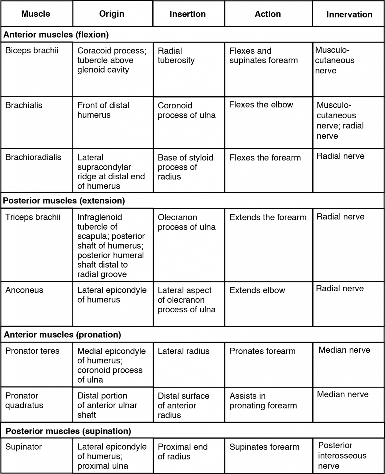
Muscles That Move the Forearm
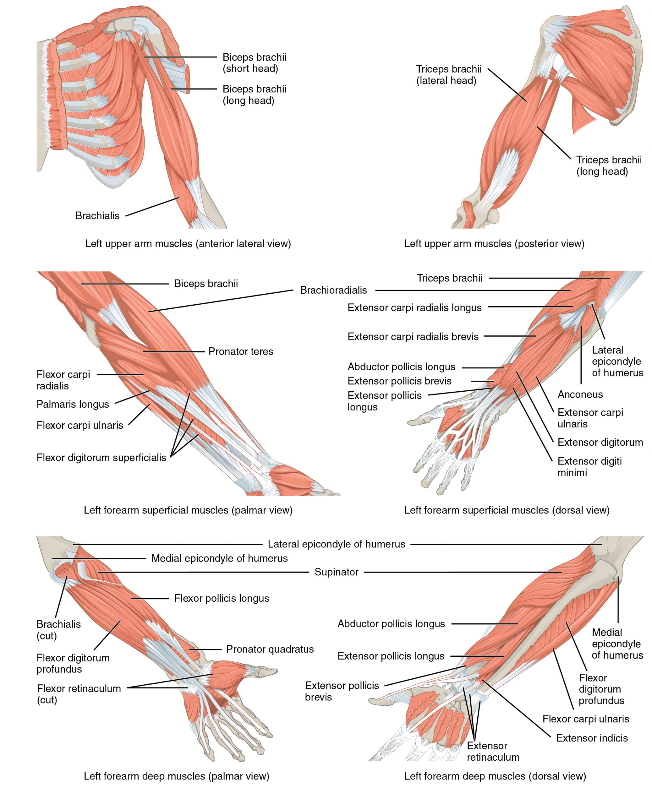
The forearm, made of the radius and ulna bones, has four main types of action at the hinge of the elbow joint: flexion, extension, pronation, and supination. When the forearm faces anteriorly, it is supinated. When the forearm faces posteriorly, it is pronated. The forearm flexors include the biceps brachii, brachialis, and brachioradialis. The extensors are the triceps brachii and anconeus. The pronators are the pronator teres and the pronator quadratus, and the supinator turns the forearm anteriorly.
The biceps brachii, brachialis, and brachioradialis flex the forearm. The two-headed biceps brachii crosses the shoulder and elbow joints to flex the forearm, also taking part in supinating the forearm at the radioulnar joints and flexing the arm at the shoulder joint. Deep to the biceps brachii, the brachial is a synergist in forearm flexion. Finally, the brachioradialis can flex the forearm quickly or help lift a load slowly. These muscles and their associated blood vessels and nerves form the anterior compartment of the arm (anterior flexor compartment of the arm) (Figure 6.4 and Figure 6.5).
Muscles That Move the Wrist, Hand, and Fingers
Wrist, hand, and finger movements are facilitated by two groups of muscles. The forearm is the origin of the extrinsic muscles of the hand. The palm is the origin of the intrinsic muscles of the hand.
Extrinsic Muscles of the Hand
The muscles in the anterior compartment of the forearm (anterior flexor compartment of the forearm) originate on the humerus and insert onto different parts of the hand. These make up the bulk of the forearm. From lateral to medial, the superficial anterior compartment of the forearm includes the flexor carpi radialis, palmaris longus, flexor carpi ulnaris, and flexor digitorum superficialis. The flexor digitorum superficialis flexes the hand as well as the digits at the knuckles, which allows for rapid finger movements, as in typing or playing a musical instrument (see Figure 6.6 and Table 11.9). However, repetitive movement with poor ergonomics can irritate the tendons of these muscles as they slide back and forth with the carpal tunnel of the anterior wrist and pinch the median nerve, which also travels through the tunnel, causing Carpal Tunnel Syndrome. The deep anterior compartment produces flexion and bends fingers to make a fist. These are the flexor pollicis longus and the flexor digitorum profundus.
The muscles in the superficial posterior compartment of the forearm (superficial posterior extensor compartment of the forearm) originate on the humerus. These are the extensor radialis longus, extensor carpi radialis brevis, extensor digitorum, extensor digiti minimi, and the extensor carpi ulnaris.
The muscles of the deep posterior compartment of the forearm originate on the radius and ulna. These include the abductor pollicis longus, extensor pollicis brevis, extensor pollicis longus, and extensor indicis (see Figure 6.6).
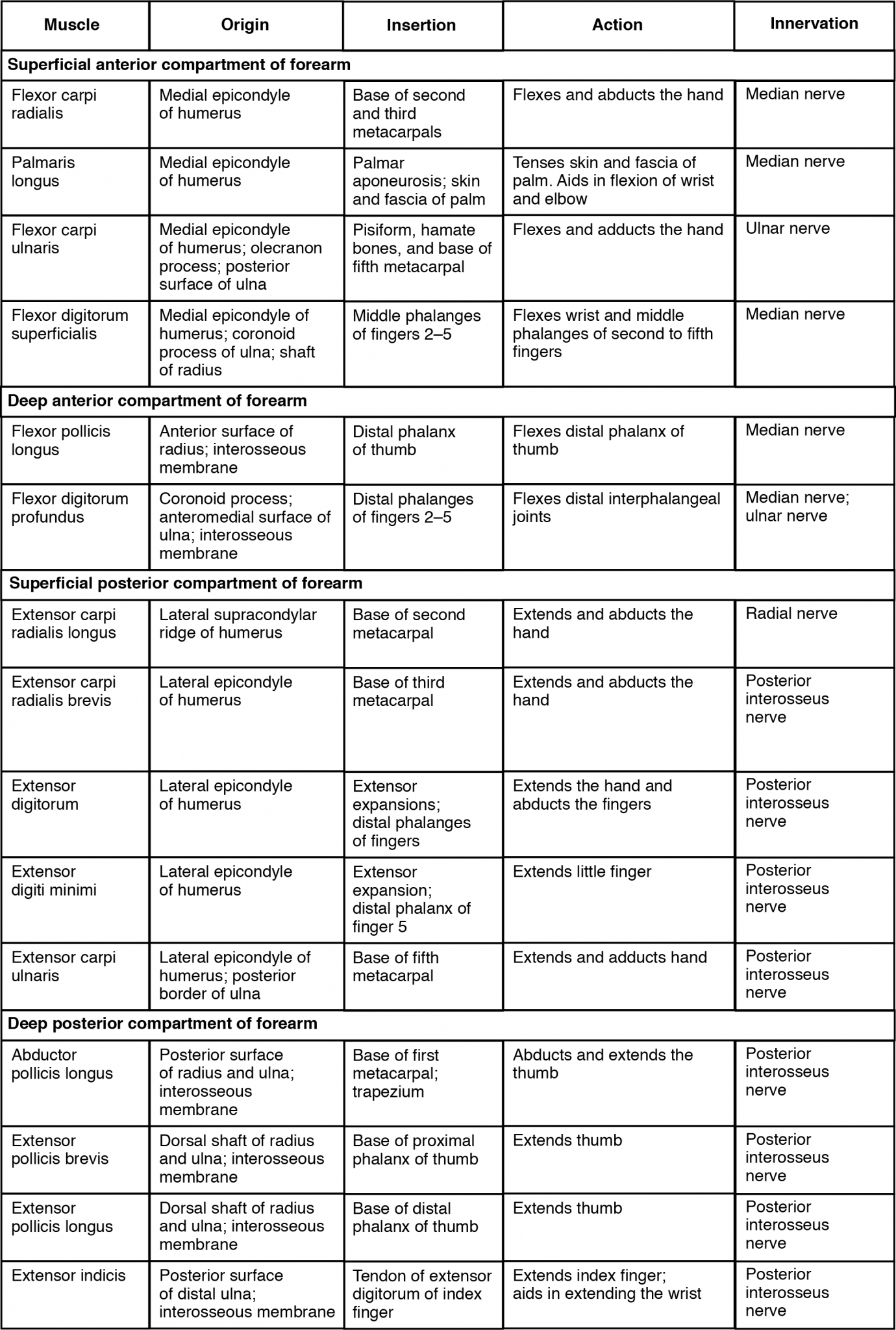
The tendons of the forearm muscles attach to the wrist and extend into the hand. Fibrous bands called retinacula sheath the tendons at the wrist. The flexor retinaculum extends over the palmar surface of the hand while the extensor retinaculum extends over the dorsal surface of the hand.
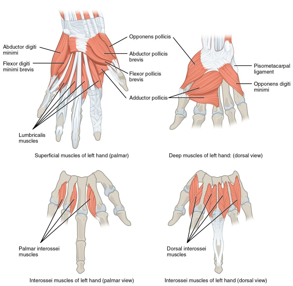
Intrinsic Muscles of the Hand
The intrinsic muscles of the hand both originate and insert within it (Figure 6.7). These muscles allow your fingers to make precise movements for actions, such as typing or writing. These muscles are divided into three groups. The thenar muscles are on the radial aspect of the palm. The hypothenar muscles are on the ulnar aspect of the palm, and the intermediate muscles are midpalmar.
The thenar muscles include the abductor pollicis brevis, opponens pollicis, flexor pollicis brevis, and the adductor pollicis. These muscles form the thenar eminence, the rounded contour of the base of the thumb, and all act on the thumb. The movements of the thumb play an integral role in most precise movements of the hand.
The hypothenar muscles include the abductor digiti minimi, flexor digiti minimi brevis, and the opponens digiti minimi. These muscles form the hypothenar eminence, the rounded contour of the little finger, and as such, they all act on the little finger. Finally, the intermediate muscles act on all the fingers and include the lumbrical, the palmar interossei, and the dorsal interossei.
| Intrinsic Muscles of the Hand (Table 11.9) | ||||||
|---|---|---|---|---|---|---|
| Muscle | Movement | Target | Target motion direction | Prime mover | Origin | Insertion |
| Thenar muscles | Moves thumb toward body | Thumb | Abduction | Abductor pollicis brevis | Flexor retinaculum; and nearby carpals | Lateral base of proximal phalanx of thumb |
| Thenar muscles | Moves thumb across palm to touch other fingers | Thumb | Opposition | Opponens pollicis | Flexor retinaculum; trapezium | Anterior of first metacarpal |
| Thenar muscles | Flexes thumb | Thumb | Flexion | Flexor pollicis brevis | Flexor retinaculum; trapezium | Lateral base of proximal phalanx of thumb |
| Thenar muscles | Moves thumb away from body | Thumb | Adduction | Adductor pollicis | Capitate bone; bases of metacarpals 2–4; front of metacarpal 3 | Medial base of proximal phalanx of thumb |
| Hypothenar muscles | Moves little finger toward body | Little finger | Abduction | Abductor digiti minimi | Pisiform bone | Medial side of proximal phalanx of little finger |
| Hypothenar muscles | Flexes little finger | Little finger | Flexion | Flexor digiti minimi brevis | Hamate bone; flexor retinaculum | Medial side of proximal phalanx of little finger |
| Hypothenar muscles | Moves little finger across palm to touch thumb | Little finger | Opposition | Opponens digiti minimi | Hamate bone; flexor retinaculum | Medial side of fifth metacarpal |
| Intermediate muscles | Flexes each finger at metacarpo-phalangeal joints; extends each finger at interphalangeal joints | Fingers | Flexion | Lumbricals | Palm (lateral sides of tendons in flexor digitorum profundus) | Fingers 2–5 (lateral edges of extensional expansions on first phalanges) |
| Intermediate muscles | Adducts and flexes each finger at metacarpo-phalangeal joints; extends each finger at interphalangeal joints | Fingers | Adduction; flexion; extension | Palmar interossei | Side of each metacarpal that faces metacarpal 3 (absent from metacarpal 3) | Extensor expansion on first phalanx of each finger (except finger 3) on side facing finger 3 |
| Intermediate muscles | Abducts and flexes the three middle fingers at metacarpo-phalangeal joints; extends the three middle fingers at interphalangeal joints | Fingers | Abduction; flexion; extension | Dorsal interossei | Sides of metacarpals | Both sides of finger 3; for each other finger, extensor expansion over first phalanx on side opposite finger 3 |